
Below are videos that makes use of gVXR. If you made your own video making use of gVXR, please let me know so that I can add it to the list.
-
Single-View Tomographic Reconstruction Using Learned Primal Dual
The Learned Primal Dual (LPD) method has shown promising results in various tomographic reconstruction modalities, particularly under challenging acquisition restrictions such as limited viewing angles or a limited number of views. We investigate the performance of LPD in a more extreme case: single-view tomographic reconstructions ...
Sean Breckling, Matthew Swan, Keith D. Tan, Derek Wingard, Brandon Baldonado, Yoohwan Kim, Ju-Yeon Jo, Evan Scott, and Jordan Pillow -
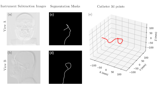
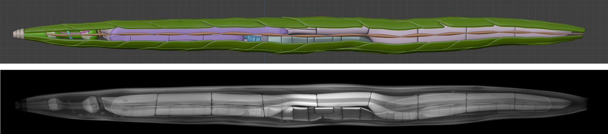
Synthetic Data Generation Pipeline for Multi-Task Deep Learning-Based Catheter 3D Reconstruction and Segmentation from Biplanar X-Ray Images
Catheter three-dimensional (3D) position reconstruction is a technology that reconstructs spatial positions from multiple two-dimensional (2D) images. It plays a pivotal role in endovascular surgical navigation, guiding surgical catheters during minimally invasive procedures within vessels. While deep learning approaches have demonstrated significant potential ...
Junang Wang, Guixiang Zhang, Wenyun Yang, Changsheng Wang, and Jinbo Yang -
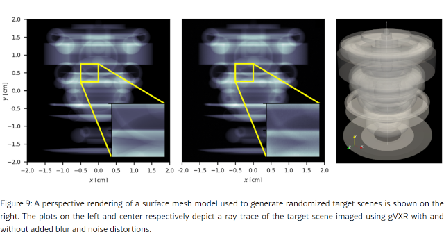
X-ray Image Generation for Robotic Radiography: a Case Study on Motion Blur in Drone-Based Wind Turbine Inspections
Robotic X-ray imaging systems enable autonomous inspection of the internal integrity of critical infrastructure. However, these systems often suffer from vibrations and unwanted movements that cause motion blur in the resulting radiographs. The impact of this motion blur is often unknown until the first prototype is available and ...
Bas Meere, Sander Doodeman, Franck P. Vidal, Paula Chanfreut, Elena Torta and Duarte Antunes -
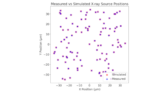
Development of an imaging protocol for laser driven X-ray sources
The Extreme Photonics Applications Centre (EPAC) being built at the Central Laser Facility in the UK will utilise a 10Hz Laser Wakefield Accelerator (LWFA) to produce a tuneable x-ray source, with energies ranging from 3keV up to 10’s of MeV while maintaining a micron-scale source size and ultra-short pulse duration. Combination of such characteristics opens an opportunity for cutting-edge high-resolution industrial imaging of dense materials... ...
E. Kiely, M. A. Williams, J. M. Warnett, K. Fedorov, O. Finlay, A. Bhardwaj, C.D. Armstrong, D. R. Symes, J. Giles-Friend and A. Bennett -
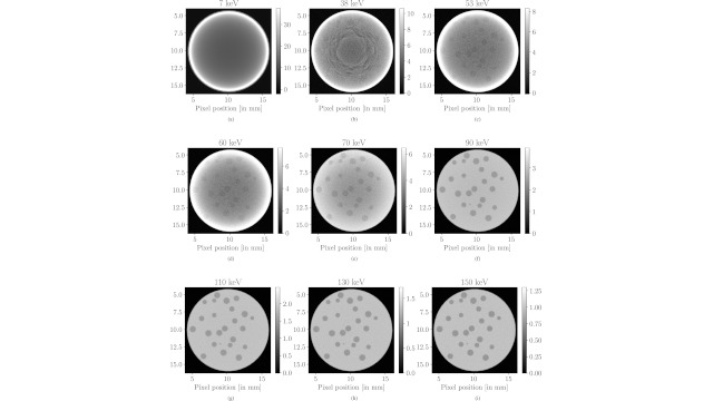
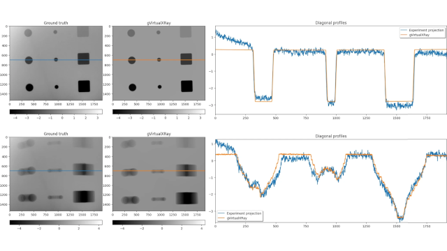
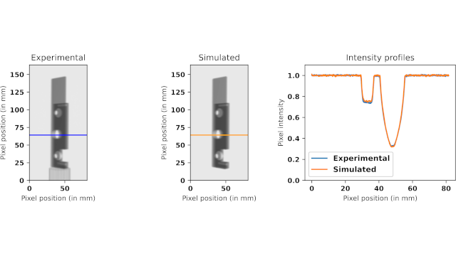
Assessing manufacturing defects in ceramic composites with both simulated and experimental synchrotron computed tomography
Non-destructive testing using X-ray computer tomography (XCT) has been used to assess the applicability of visualising ceramic kernels held within a dissimilar ceramic matrix. Simulations were performed to ascertain the feasibility of CT scans of such samples, and optimise the scanning parameters offline. ...
Iwan T. Mitchell, Fabio Martini, Gareth F. Stephens, Simon C. Middleburgh and Franck P. Vidal -
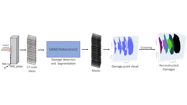
Investigation of deep learning approaches for automated damage diagnostics in fiber metal laminates using Detectron2 and SAM
The impact damage is one of the major causes of structural failures in Fiber Metal Laminate (FML) plates, which are widely used in the aerospace and automotive industries due to their superior mechanical properties. Accurate detection, segmentation, and characterization of these damages are crucial for improved safety and reduced maintenance costs...
Sanjeev Kumar, Stefan Bosse, and ShahChirag Shah -
Additive manufacturing in aluminium of a primary mirror for a CubeSat application: manufacture, testing, and evaluation
Additive manufacturing (AM; 3D Printing), a process which creates a part layer-by-layer, has the potential to improve upon conventional lightweight mirror manufacturing techniques, including subtractive (milling), formative (casting) and fabricative (bonding) manufacturing. Increased mass reduction whilst maintaining mechanical performance can be achieved through the creation of intricate lattice geometries ...
Ilhan Aziz et al. -
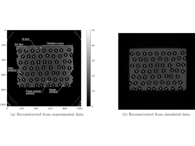
Hybrid Modeling of Material Imperfection Indications in X-ray Computed Tomography Datasets
Machine learning, more specifically Deep Learning models, have shown remarkable potential for analyzing 3D CT data within both material science and NDT applications. However, the models require large amounts of training data, especially Deep Learning models. Because high quality real experimental data is expensive to derive, humans annotating, synthetic training data is often utilized. In this work we have explored if training data can be modeled with a hybrid approach; ...
Erik Lindgren, Fabian Hanning, and Linda Squillaci -

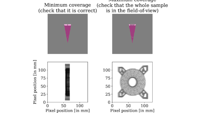
WebCT - OpenSource Web-based GUI for Realtime X-ray Simulation
Scan planning for X-ray CT systems can be difficult due to the large number of elements affecting scan quality. The use of X-ray simulation can answer feasibility questions, however existing methods are focused on experts who are familiar with XCT and programming knowledge. WebCT is a user-centric application for performing virtual XCT scans with the validated X-ray simulator gVirtualXray. Focused on accessibility, the interface ...
Iwan T. Mitchell, Jean Michel Létang, Llion M. Evans and Franck P. Vidal -
Developing next-generation algorithms for X-ray emission micro-tomography
X-ray emission micro-imaging, whether induced by protons or synchrotron X-rays (SXRF: Synchrotron X-Ray Fluorescence), enables multi-scale mapping of biologically relevant elements, such as minerals and trace metals, with high mass sensitivity. ...
A. L. Brun, P. Desbarats, J.-F. Giovannelli, C. Michelet, A. Rouwane and H. Seznec -
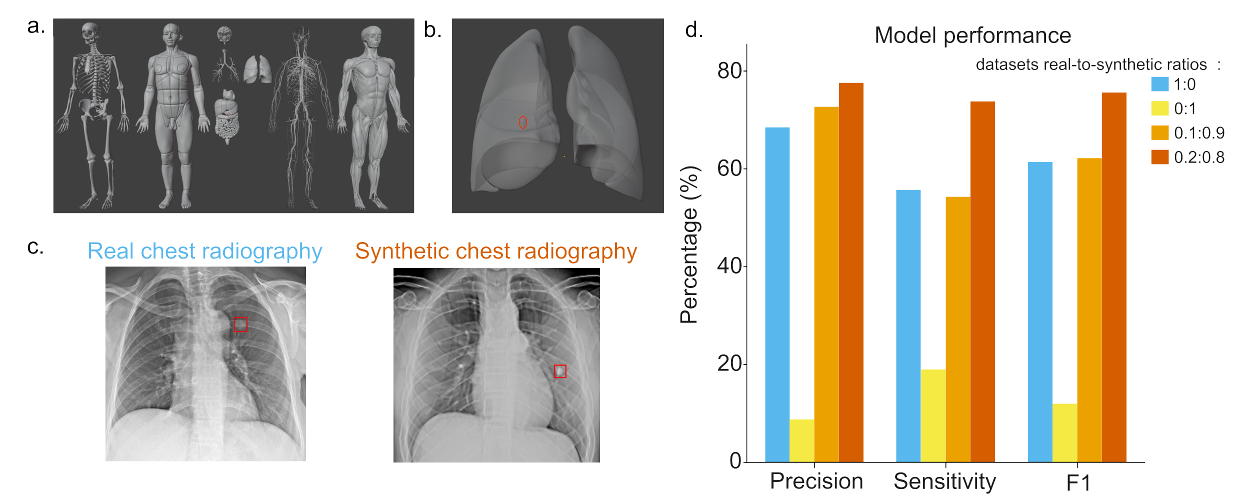
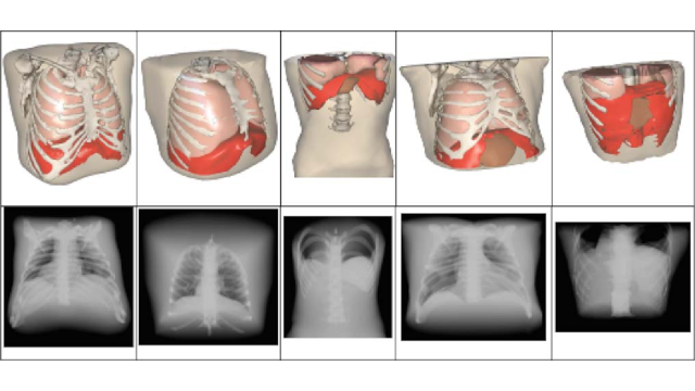
Lung nodule detection in 2D radiographs
Deep learning models are a promising approach to enhance medical image analysis. However, their performance is limited by the size, quality, and diversity of available training datasets. Incorporating synthetic data into training datasets can address the lack of medical imaging data. The combined use of a virtual anthropomorphic model with gVirtualXRay enables the rapid generation of large synthetic datasets. ...
Clémentine Hatton and Audrey Henry -
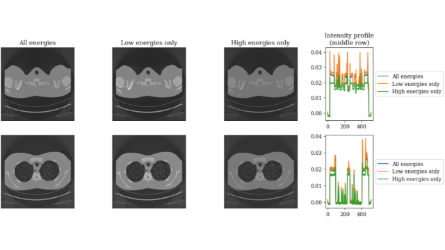
Spectral CT data simulation and material decomposition
The ability of gVirtualXRay to simulate spectral CT data plays a crucial role in advancing material decomposition techniques within medical imaging. By generating synthetic datasets across high-energy, low-energy, and conventional CT spectra, gVXR provides essential training data for deep learning models in material decomposition. ...
Shaghayegh Afshari and Cheng-Ying Chou -
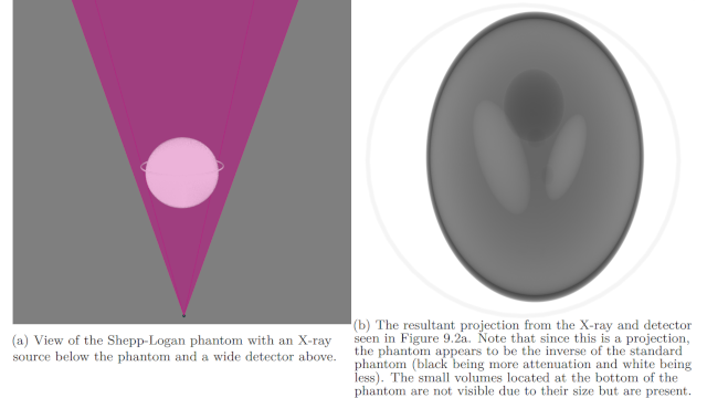
Physics Integrated Deep Learning For Photon Counting Spectral CT (Doctoral dissertation), Chapter 9 Mars V6 Simulator
This chapter describes the implementation of a simulator for the MARS V6 environment. The purpose of creating a simulator was to aid in debugging and implementing the reconstruction approach. ...
Aaron Smith -
Hybrid Modeling of Material Imperfection Indications in X-ray Computed Tomography Datasets
This article presents a fluoroscopy image-based registration method along with a comprehensive protocol for robotic needle insertion in radiofrequency ablation (RFA) to treat liver cancer. ...
Thuc-Long Ha, Juan Verde, Julien Bert, and Hadrien Courtecuisse -
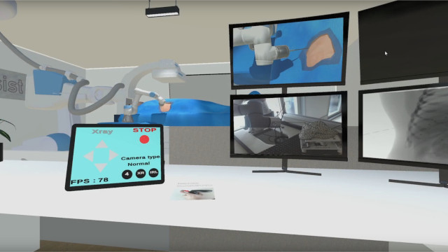
Mixed Reality and Real-Time X-Ray Simulation in Vet Radiography Training: A User-Centered Comparative Study
Proficient training in Diagnostic Imaging (DI) is crucial for veterinary students. However, the training process faces challenges, including limited access to animals and strict radiation safety regulations. ...
Xuanhui Xu, Antonella Puggioni, David Kilroy, and Abraham G. Campbell}, -
Teaching XCT in NDT using Simulation on GPU with gVirtualXray
We have deployed gVXR in material science lab-sessions delivered to about 150 MEng students a year at the INSA-Lyon and Polytech Lyon 1 engineer schools. It has been embedded in a Jupyter notebook together with the open-source reconstruction toolkit RTK. Several interactive exercises have been proposed to enable students to learn and gain hands-on experience with X-ray tomographic setups. ...
Jean Michel Létang and Franck P. Vidal -
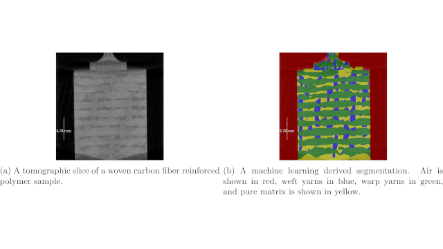
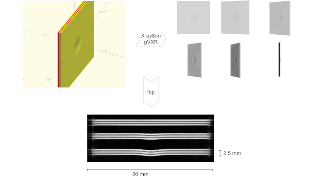
Automated generation of labeled synthetic training data for machine learning based segmentation of 3D-woven composites
Composite parts with 3D-textile reinforcement show promise in high-performance applications. For widespread use, accurate material characterisations are required. Characterisation of the textile architecture in the as-manufactured state may be performed with X-ray CT. ...
Johan Friemann, Lars P. Mikkelsen, Carolyn Oddy and Martin Fagerström -
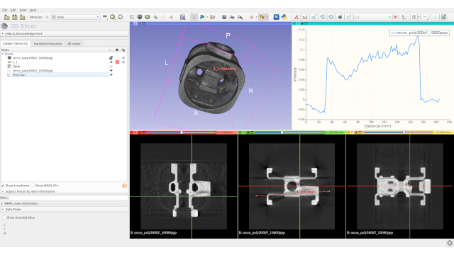
How does a manufactured object compare to its original CAD design?
We investigate methods of assessing how a manufactured part differs from a reference CAD model. We use a part of an in-situ tensile testing machine designed to be used with a scanning electron microscope. CAD drawings are available, and the corresponding 3D surface is exported using the STL file format. After manufacturing, a CT scan was acquired with the DTHE scanner ...
Franck P. Vidal, Iwan T. Mitchell, Jean-Yves Buffière and Jean Michel Létang -
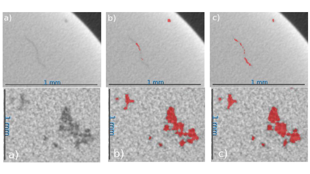
Automated Porosity Characterization for Aluminum Die Casting Materials Using X-ray Radiography, Synthetic X-ray Data Augmentation by Simulation, and Machine Learning
Detection and characterization of hidden defects, impurities, and damages in homogeneous materials like aluminum die casting materials, as well as composite materials like Fiber–Metal Laminates (FML), is still a challenge. ...
by Stefan Bosse, Dirk Lehmhus and Sanjeev Kumar -
XRayViewer - A C++ Unreal Engine 5 Wrapper for gVirtualXRay
Lab-CT is a rapidly developing area of research. As CT scanners have become more compact and cheaper, they are more accessible to researchers. This however introduces the problem of training users to correctly operate lab-CT scanners. It is often difficult or not practical to allow researchers time to train thoroughly on equipment before they use it, as this takes up precious time allocated to the researchers. With this in mind, XRayViewer aims to be a generic training tool for researchers to use to gain valuable experience with ...
Lewis Dixon -
Use of fast realistic simulations on GPU to extract CAD models from microtomographic data in the presence of strong CT artefacts
The presence of strong imaging artefacts in microtomographic X-ray data makes the CAD modelling process difficult to carry out. As an alternative to traditional image segmentation techniques, we propose to register the CAD models by deploying a realistic X-ray simulation on GPU in an optimisation framework. A user study was also conducted to compare the measurements made manually by a cohort of ...
Franck P. Vidal and Iwan T. Mitchell and Jean M. Létang -
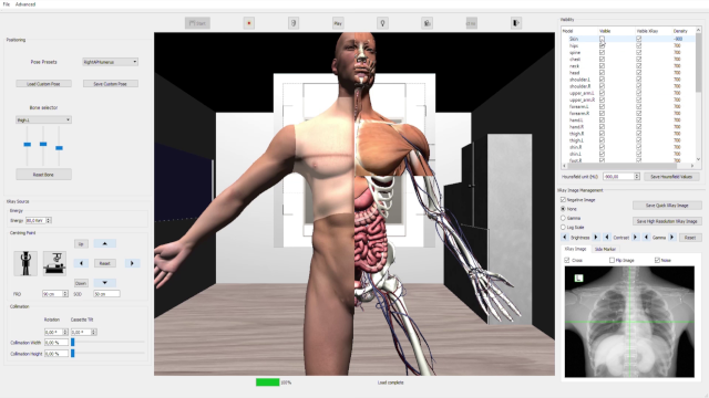
Interactive teaching environment for diagnostic radiography with real-time X-ray simulation and patient positioning
Traditional undergraduate radiographer training mixes academic lectures and clinical practice. Our goal is to bridge the current disconnection between theory and practice in a safe environment, avoiding the risk of radiation for both practitioners and patients. To this end, this research proposes a new software to teach diagnostic radiography using real-time interactive X-ray simulation and patient positioning.
Aaron Sujar, Graham Kelly, Marcos Garcia, and Franck P. Vidal -
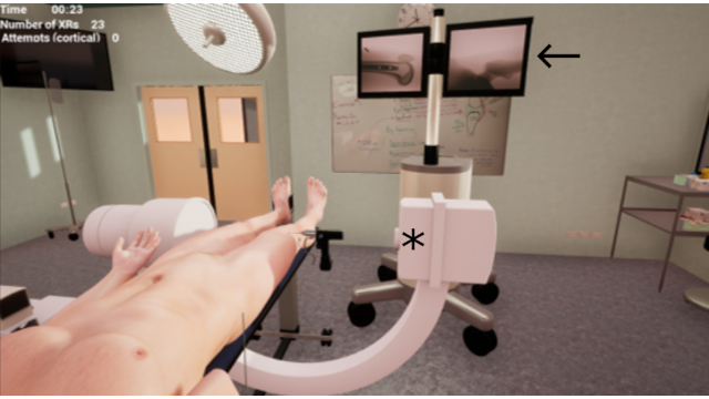
Development and Validation of a Virtual Reality Haptic Femoral Nailing Simulator
The simulator consisted of a 3D virtual environment, a haptic device and 3D printed drill handle and a VR headset. The environment was created using a video game development engine, interfaced with plugins to allow haptic feedback and image intensifier functionality. Two tasks were created within the simulator as part of an antegrade femoral intramedullary (IM) nail procedure: proximal guidewire entry and distal locking.For the validation study, participants performed the above tasks on the simulator. ...
Malek Racy, Alastair Barrow, James Tomlinson and Fernando Bello -

Registration of a generic 3D hand model on 2D radiographs using x-ray simulation and CMA-ES
Radiographs of the hand are useful in diagnosing and staging diseases such as rheumatoid arthritis (RA) and other musculoskeletal diseases. Radiographs are projections of the 3D anatomy, with the useful information such as pose and pathology becoming lost in the process. We propose a 3D hand pose recovery method for radiographs of hands using a novel hybrid image registration method. Our pose recovery pipeline consists of aligning a simulated X-ray ...
Tianci Wen and Radu P. Mihail, and Franck P. Vidal -

X-ray imaging virtual online laboratory for engineering undergraduates
Distance learning engineering students (as well as those in face-to-face settings) should acquire a basic background in radiation–matter interaction physics (usually in the first semesters). Some students in this group may feel some degree of aversion towards these types of pure science-related subjects (mathematics, physics, chemistry, etc). In online learning scenarios, the average student is already an adult (37 years old or above) and ...
Alberto Corbi, Daniel Burgos and Franck P. Vidal, Francisco Albiol and Alberto Albiol -
Densitometric radiographic imaging with contour sensors
We present the technical/physical foundations of a new imaging technique that combines ordinary radiographic information (generated by conventional X-ray settings) with the patient's volume to derive densitometric images. Traditionally, these images provide quantitative information about tissues densities. In our approach, they graphically enhance either soft or bony regions. After measuring the patient's volume with contour recognition devices, ...
Francisco Albiol, Alberto Corbi and Alberto Albiol -
Simulated motion artefact in computed tomography
We propose a simulation framework to simulate the computed tomography acquisition process. It includes five components: anatomic data, respiration modelling, automatic parametrisation, X-ray simulation, and tomography reconstruction. It is used to generate motion artefacts in reconstructed CT volumes. Our framework can be used to evaluate CT reconstruction algorithm with motion artefact correction in a controlled environment.
Franck P. Vidal and Pierre-Frédéric Villard -
A prototype percutaneous transhepatic cholangiography training simulator with real-time breathing motion
We present here a simulator for interventional radiology focusing on percutaneous transhepatic cholangiography (PTC). This procedure consists of inserting a needle into the biliary tree using fluoroscopy for guidance. ...
Pierre-Frédéric Villard, Franck P. Vidal, Carrie Hunt, Fernando Bello, Nigel W. John, Sheena Johnson and Derek A. Gould