
Diamond DIAD
Overview
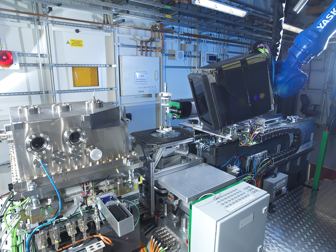
The DIAD (Dual Imaging and Diffraction) beamline at Diamond Light Source is a unique synchrotron instrument designed to acquire imaging and diffraction1 measurements from the same specimen location, quasi-simultaneously.
1 diffraction is not part of the twin in gVXR.
Beam Characteristics
| Parameter | Value |
|---|---|
| Energy range | 7–38 keV |
| Geometry | Parallel beam |
| Acquisition | Imaging + diffraction |
| Beam type | Monochromatic / pink beam |
Scientific Applications
The DIAD beamline is particularly suited to research involving:
- Battery materials
- Additive manufacturing
- Metals and alloys
- Phase transformations
- Residual stress analysis
- Catalysis
- Geological materials
- Biomaterials
- In-situ and operando experiments
The unique dual-beam design allows tomography and diffraction1 measurements to be acquired quasi-simultaneously at the same location within a specimen, enabling direct correlation between morphology and crystallographic information.
Twin Fidelity
Unlike a purely geometrical representation, the DIAD digital twin includes experimentally measured and calibrated parameters:
- Measured synchrotron spectra
- Measured detector line-spread function (LSF)
- Calibrated flux curves
- Detector gain model
- Dark-field response
- Scintillator energy response
- Mechanical positioning constraints
These models enable realistic simulation of image quality, noise, blur, and beam hardening effects observed on the real instrument.
This allows realistic simulation of DIAD experiments before beamtime and supports reconstruction development, machine-learning dataset generation, and scan protocol optimisation.
X-ray Source
Monochromatic Beam
| Property | Value |
|---|---|
| Geometry | Parallel beam |
| Energy range | 14.01–37.92 keV |
| Flux model | Energy-dependent calibrated flux |
| Detector blur | Measured LSF included |
Pink Beam (Low Angle)
| Property | Value |
|---|---|
| Geometry | Parallel beam |
| Spectrum | Measured spectrum |
| Energy range | ~1.5–49.5 keV |
| Filtration | Al (0.1–4 mm), Cu (0.5 mm) |
Pink Beam (High Angle)
| Property | Value |
|---|---|
| Geometry | Parallel beam |
| Spectrum | Measured spectrum |
| Energy range | ~1.5–49.5 keV |
| Filtration | Al (0.1–4 mm), Cu (0.5 mm) |
Detector Model
The digital twin includes a physics-based detector model calibrated from experimental measurements.
Detector Characteristics
| Property | Value |
|---|---|
| Resolution | 2560 × 2160 pixels |
| Pixel pitch | 0.5 μm |
| Scintillator | Gd₃Ga₅O₁₂ |
| Scintillator thickness | 170 μm |
| Dark-field noise | Included |
| Detector gain | Included |
| Line spread function | Measured and modelled |
Simulated Effects
- Detector blur (LSF)
- Detector gain
- Scintillator energy response
- Dark-current bias
- Dark-current noise
- Quantum noise through calibrated flux measurements
Acquisition Geometry
Source Position
| Parameter | Value |
|---|---|
| Source-to-object distance | 47.5 m |
Detector Position
| Parameter | Value |
|---|---|
| Object-to-detector distance | 1.25 mm – 1.37 m |
| Typical maximum used experimentally | ~300 mm |
This geometry enables both absorption-contrast and propagation-based phase-contrast imaging.
Why This Twin Is Unique
The DIAD digital twin reproduces one of the world’s few beamlines designed for quasi-simultaneous imaging and diffraction measurements on the same specimen.
The implementation goes beyond simple geometry reproduction by incorporating experimentally measured spectra, calibrated flux models, detector-response characteristics, detector blur measurements, and mechanical constraints observed on the real instrument.
This enables realistic simulation of synchrotron imaging experiments and provides a valuable platform for experiment planning, reconstruction development, and machine-learning dataset generation before beamtime.
The twin is particularly valuable for researchers interested in correlating microstructural imaging with diffraction-based material characterisation.
Affiliations
- Facility: Diamond Light Source
- Beamline: DIAD - Dual Imaging and Diffraction
- Country: United Kingdom